Overview
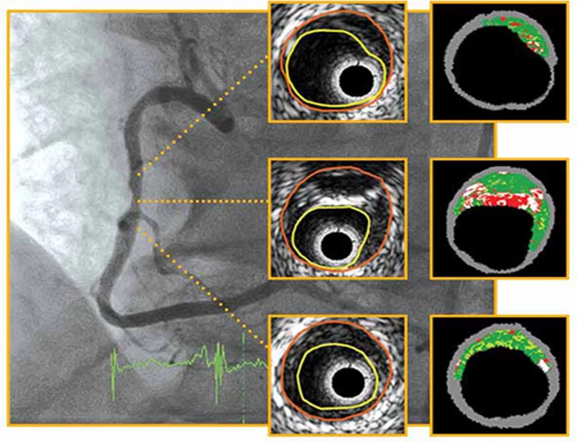
An angiogram shows the outline of the artery like a shadow. Intravascular ultrasound (IVUS) and optical coherence tomography (OCT) use a tiny camera-like probe inside the artery to show the vessel wall itself.
They reveal how much plaque is present, whether it is soft or calcified, and the true size of the artery — so the right stent can be chosen and fully expanded. This lowers the chance of the artery narrowing again.
Who may need it
- Complex blockages: left main artery, long lesions, bifurcations
- Heavily calcified arteries
- Checking an old stent that has narrowed again
What to expect
- Done during angiography or angioplasty through the same catheter.
- The imaging probe is advanced past the blockage and slowly pulled back while recording.
- It adds only a few minutes to the procedure.
Recovery & aftercare
- Recovery is the same as for the angiogram or angioplasty it is part of.
Medically reviewed by Dr. G.V. Senthilnathan, MBBS, MD (General Medicine), DM (Cardiology). This page is general information; your treatment plan will be tailored to your own reports and health.